|
|
|
|
In the 1990s, researchers at Varian's Ginzton Technology Center in Palo Alto, California developed a digital replacement for X-ray film and image intensifier tubes used in diagnostic imaging. The final product - amorphous silicon (a-Si) flat panel systems capable of both fluoroscopic and radiographic image acquisition on the same receptor - combines speed, image quality, compactness and ease of use. Delivered first by Varian, more than 100 of these systems are now being used in diverse application areas from neonatal intensive care units to steel mills. 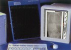 Varian's PaxScan amorphous silicon image receptors could take the place of medical X-ray film, providing instant diagnostic images, reducing patients' exposure to radiation, and eliminating the need for film processing and storage.
|
 Very premature babies desperately need oxygen, nourishment, and other life support.The trick is precisely placing tubes and support lines into the tiny lungs and blood vessels of patients no bigger than your hand.
In most hospitals, X-ray films are taken to verify placement. But taking and developing film is a cumbersome, time-consuming process, especially when the radiology department is located several floors away from the neonatal intensive care unit (NNICU). In late 1999, Varian's X-Ray Products business installed a PaxScan 2520 amorphous silicon (a-Si) flat panel image receptor in the Pediatric Radiology Department at the Medical University of South Carolina (MUSC). The panel system is being tested for potential utility in radiological evaluation of premature infants.
"Nurses sometimes must sit still for as long as an hour, holding a line in a premature infant with a collapsed lung or other condition requiring immediate treatment, while they wait for X-ray results to come back from radiology," says Dr. William Michael Southgate, Director of the NNICU Department at MUSC and collaborator in the study. Varian's receptor is placed directly under the infant. A portable X-ray unit is used to make an exposure. The images are displayed instantly on a monitor. In this way, doctors can adjust support lines within minutes. Dr. Hill says the flat panel system needs only one-third the radiation required by X-ray film. "These preemies have dozens of X-rays in the first years of their lives. We must remain concerned about cumulative radiation dose, and the a-Si flat panel technology relieves this concern." |
|
|